
It turns out that psychopaths' brains are very different.

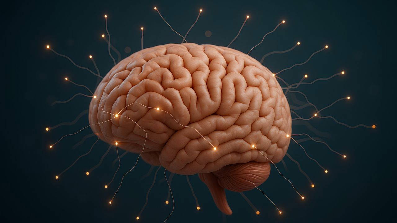
Significant structural differences have been found in the brains of individuals with high levels of psychopathic traits in regions associated with impulse control and emotional regulation. This new study, conducted jointly by researchers from the US and Germany, offers important insights into better understanding these individuals and may also lay the scientific groundwork for future rehabilitation approaches.
Psychopathy is one of the most misinformed concepts in society. While often used synonymously with "evil," modern psychiatric manuals lack a formal diagnostic category for "psychopathy." Instead, traits such as a lack of emotion, manipulative attitudes, superficial charm, lack of empathy, and antisocial behavior are considered personality patterns and presented on a spectrum.
Individuals with a more pronounced personality trait, as seen in Chip, are known to be more likely to exhibit violent behavior, criminal activity, and recidivism. Therefore, exploring the relationship between psychopathic traits and the brain is considered a topic with significant implications for both the individual and the societal level.
Differences in the brainIn the new study, the brains of 39 male participants with high psychopathy scores were examined using functional MRI imaging. The participants were assessed using the Psychopathy Checklist-Revised (PCL-R), a 20-item scale widely used in clinical studies that measures two core dimensions of psychopathy: the first dimension encompasses emotional detachment and aloofness, and the second dimension encompasses behaviors related to antisocial tendencies.
Individuals high in psychopathy were compared with a control group without psychopathic traits. Volumetric analyses using the Julich Brain Atlas revealed significant structural differences, particularly in regions corresponding to the second dimension (antisocial behavior).
The study revealed volume loss in regions such as the basal ganglia, thalamus, and insular cortex. These regions are associated with many fundamental functions, including impulse control, social cognition, reward perception, and emotional processing. Overall, individuals with high levels of psychopathy had approximately 1.45 percent less volume in their brains compared to controls. The most striking differences were concentrated in certain parts of the cortex, the anterior cingulate, and specific subfields of the hippocampal formation.
What do the findings mean?The results suggest that behaviors associated with psychopathy may not be explained solely by social or environmental factors; they may also have a neurological basis. The research team speculates that psychopathic tendencies may emerge as a developmental brain difference in some individuals.
However, the study also has limitations. The relatively small sample size raises some questions about the generalizability of the results. Furthermore, although it is assumed that the participants were not under the influence of substances at the time of the study, long-term substance use in the past may have affected brain structure. This can make the data somewhat difficult to interpret.
The researchers emphasize the need for more comprehensive neuroimaging studies on psychopathy. Such data are crucial for both early detection of individuals and the development of appropriate intervention strategies.
The study was published in the journal European Archives of Psychiatry and Clinical Neuroscience.
Cumhuriyet


